Medical Image Processing
Digital processing of medical images poses an ongoing challenge for computer science. The required accuracy and reliability in medical systems impose high technical and mathematical demands. Increased complexity and the growing amount of data motivate investigations in the field of knowledge-based image analysis where semantic models for object recognition are developed.
Registration
Registration of medical images from different modalities, viewing directions, times and dimensions requires the application of complex mathematical methods to compensate for system immanent deformations and the lack of exact correspondences.
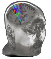
Image Fusion
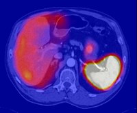
Fusion of images from different acquisition methods, like X-Ray, CT, MRI, PET or SPEC, is a key aspect for diagnosis as well as for the planning of surgical interventions. Providing meaningful visualizations from multiple sources requires accurate registration techniques and powerful interactive displaying methods.
Segmentation (2D, 3D)
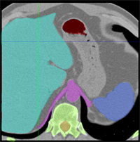
Medical image segmentation is the process of partitioning data into regions, which often correspond to different tissue classes, organ pathologies, or other anatomically relevant structures. Typically, medical images exhibit low contrast or have a high degree of noise which makes segmentation difficult. Many computer vision techniques for segmentation have been adapted specifically for medical image modalities.
Biomechanical Modelling
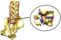
MultiBody-Simulation (MBS) is a modern method to determine stress and strain in biological structures of the human body. Load changes of internal structures and the change of kinematics during different external forces can be analyzed. With our MBS model of the lumbar spine it is possible to calculate the inner forces and torques occurring in the specific lumbar structures and to determine the resulting kinematics. Furthermore, mechanical effects of interventions to adjacent vertebral segments can be estimated.
© Universität Koblenz